2023 Research Grants
We are delighted to announce the AMRF grant recipients for 2023.
Melanoma incidence is set to rise over the next 10 years. More than 200,000 Australians are expected to be diagnosed with melanoma before 2030.
The survival rate from advanced melanoma has improved from 10% to 50% over the last decade – largely due to improvements in detection and treatment.
It is only through the support of our partners and donors that this support is available. Thank you for making a difference. Our Research Committee, which includes two external experts, has selected five young Australian researchers for grants in the 2023 round.
We are also delighted to announce the recipients of the Warren Meanwell Melanoma Research Grant and the LEK Consulting Melanoma Research Grant for 2023.
Early Career Research Scientist and Warren Meanwell Melanoma Research Grant recipient for 2023
Identifying the gene expression signatures of tumour-specific CD8+ T cells in adjuvant anti-PD-1 treated stage III melanoma patients
Grace Attrill
The University of Sydney, NSW
Early Career Research Scientist and LEK Consulting Research Grant recipient for 2023
Research into the identification of protein signatures and the use of scarless skin biopsies to achieve more reliable diagnoses through proteomic analysis
Dr Rachel Teh
The University of Sydney, NSW
Early Career Research Scientist Grant recipient for 2023
IgG and IgA autoanti-bodies as predictive biomarkers of immune-related adverse events (irAEs) and survival in cutaneous melanoma patients on immune checkpoint inhibitors
Dr Pauline Zaenker
Edith Cowan University, Perth, WA
Early Career Research Scientist Grant recipient for 2023
Assessing behavioural impacts of receiving personalised risk scores for melanoma
Dr Tatiane Yanes
The University of Queensland, QLD
Post Graduate Research Grant recipient for 2023
Immunosuppression and the tumour microenvironment in advanced melanoma
Catherine Zilberg
The University of Sydney, NSW

“I feel very fortunate to receive the Warren Meanwell Melanoma Research Grant for 2023 for this exciting new study, which we hope will lead to more effective therapies for melanoma patients. Thank you very much!”
Grace Attrill
Identifying the gene expression signatures of tumour-specific CD8+ T cells in adjuvant anti-PD-1 treated stage III melanoma patients
Grace Attrill
The University of Sydney, NSW
CD8+ T cells are immune cells which can target and destroy cancer cells, and many immune therapies – such as anti-PD-1 therapy – work by boosting these cells. Immunotherapy is not 100% effective for all melanoma patients, and researchers are currently working to figure out why. Intriguing new research has suggested that many of the CD8+ T cells in tumours do not target melanoma cells. Understanding how this occurs could be vital to improving melanoma therapies and reducing treatment side-effects. However, with current technologies it’s difficult to differentiate between the CD8+ T cells that target melanoma – the ‘melanoma-specifics’ – and those that do not – the ‘bystanders’.
Our study will combine multiple cutting-edge technologies to generate gene expression signatures that distinguish melanoma-specific and bystander CD8+ T cells in the tumours and blood of metastatic melanoma patients treated with anti-PD-1 therapy. With these signatures, we will learn more about melanoma-specific and bystander CD8+ T cell functions and their roles in immunotherapy response and resistance. It is hoped that generating these CD8+ T cell profiles for melanoma patients will lead to more personalised and effective therapies.
July 2024 update
My project involves the gene expression profiling of CD8+ T cells based on their ability to recognise and destroy melanoma cells. Over the past 12 months, we have optimised and completed an experimental protocol to study this unique population of cells in the blood and tumours of melanoma patients. By combining dextramer staining and FACS-based cell sorting, we isolated melanoma-specific and bystander CD8+ T cell populations. We then performed single-cell RNA and TCR sequencing on these isolated CD8+ T cells.
All experimental work and data collection for this project is now complete, with CD8+ T cells isolated from the tumours and blood of 9 metastatic melanoma patients. We can now analyse the expression of every single gene in each cell, which will allow us to assess multiple characteristics of these CD8+ T cells including their role in immunotherapy response. With the collected TCRseq data, we will also be able to test which CD8+ T cell populations have expanded in response to the tumour, as well as track the migration of CD8+ T cell clones from the blood to the tumour. Combined, this is a substantial amount of data, and we are now undertaking the complex and extensive work required to analyse it.
I am looking forwards to completing this work, and to sharing our findings with the AMRF. Thank you again for your support!
Research into the identification of protein signatures and the use of scarless skin biopsies to achieve more reliable diagnoses through proteomic analysis
Dr Rachel Teh
The University of Sydney, NSW
My research involves investigating the utility of a scarless biopsy technique which collects skin samples using adhesive tape. Proteins are extracted from the collected skin samples to identify a protein signature which will diagnose melanoma in suspicious moles. We are recruiting patients undergoing full body photography through the Australian Centre of Excellence in Melanoma Imaging and Diagnosis (ACEMID) to provide comprehensive imaging and histopathology together with the scarless biopsy.
We aim to develop a diagnostic approach which will reduce the number of removed moles with no malignant potential, improve detection of melanoma, and provide an accessible tool for patients in rural and remote communities.

“I’m grateful and honoured to be awarded by the AMRF. This means that I can continue my research towards improving the way we diagnose melanoma and develop a diagnostic tool which will help underserved communities. Thank you, AMRF.”
Dr Rachel Teh
July 2024 update
I am looking into the utility of a scarless biopsy technique to diagnose melanoma. This involves the collection of skin samples using tape. We hope to develop a diagnostic approach which will reduce the number of benign moles being removed whilst improving detection of melanoma and provide an accessible tool for patients in rural and remote communities.
By recruiting patients undergoing full body photography through The Australian centre of Excellence in Melanoma Imaging and Diagnosis (ACEMID) projects, we have collected 271 scarless samples with matching dermoscopy and histopathology. Our cohort currently consists of samples diagnosed as benign naevi n=96, dysplastic naevi n=136, melanoma in situ n=7, and invasive melanoma n=2. With the help of Princess Alexandra Hospital (QLD), and Alfred Health (Vic) we will continue to acquire our targeted melanoma samples and commence analysis for biomarkers to validate on our way to develop the scarless biopsy as a diagnostic tool for melanoma. I am grateful for the Australian Melanoma Research Foundation and LEK Consulting Award in supporting my work.
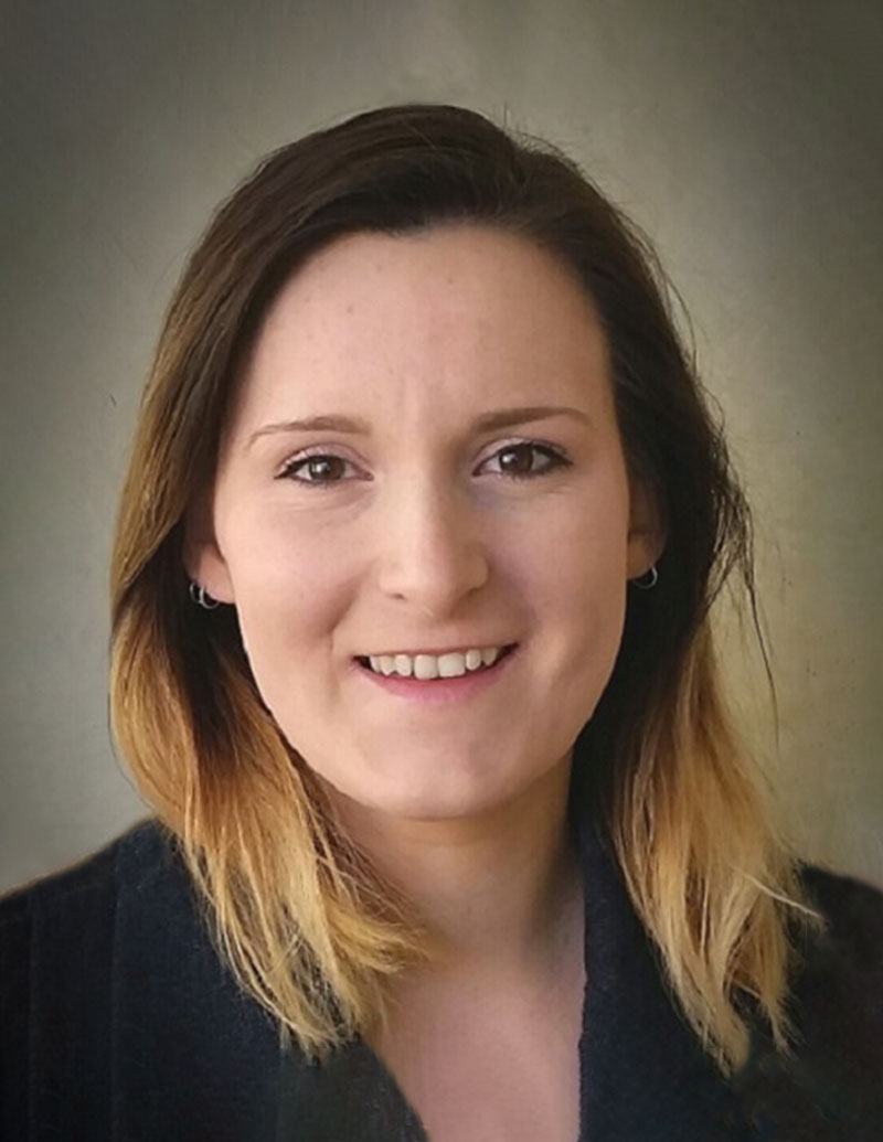
“As an early career researcher, I am passionate about the development of tests that can serve as additional tools to improve the diagnostic and prognostic certainty of cancer surveillance. I specialise in the clinical application of next generation proteomics and am driven by an interest in research translation and articulating scientific knowledge to the public. The AMRF Early Career Research Scientist grant enables me to validate research findings to further the translation of my research into the clinic.”
Dr Pauline Zaenker
IgG and IgA autoanti-bodies as predictive biomarkers of immune-related adverse events (irAEs) and survival in cutaneous melanoma patients on immune checkpoint inhibitors.
Dr Pauline Zaenker
Edith Cowan University, Perth, WA
The use of immunotherapy has revolutionised the melanoma treatment landscape in the last decade. End-stage melanoma patients now experience improved survival, but many suffer from debilitating, potentially severe grade immune-related side adverse events (irAEs) that require hospitalisation, potential treatment disruption and may be fatal. Biomarkers in the blood of patients that can predict treatment-related irAEs do not currently exist but are critical for the monitoring and prevention of these severe side effects. Based on our previous data, we believe that autoantibodies, markers of the immune system, may serve as predictive biomarkers of severe side effects.
We recently identified and will now validate a shortlist of autoantibodies that may be predictive of severe side effects and these may prove valuable for the development of a blood test that can improve the prediction and early detection of immunotherapy side effects in melanoma patients. In the future, such a blood test has the potential to aid in clinical decision-making with regard to treatment options. This will especially be important in coming years since these treatments are now considered for use in much earlier tumour stages (stage 2 onwards) and the predicted risk of irAE development will be an important consideration for the use of these treatments at early disease stages.
July 2024 update
We are pleased to share the progress on our research project, which focuses on validating biomarkers for irAE prediction in melanoma patients undergoing ICI treatment. For Aim 1, we are using a focused microarray to validate IgA and IgG antibody levels in a subset of our original patient cohort. Aim 2 involves validating these antibodies in an independent cohort of over 50 stage IV melanoma patients, while Aim 3 includes ongoing pathway analysis using Advaita Bio’s ipathwayguide software to understand the biological significance of our biomarkers.
Since the grant was awarded, we have acquired an InnoScan 710 microarray scanner with Mapix software through internal funds, enabling in-house validation. We continue to collaborate with Dr. Yu (Curtin University, WA), and our pathway analysis of our initial discovery study confirmed the biological relevance of identified biomarkers in melanoma and immune responses.
We have discussed these results with our research team and during consumer meetings, completed the selection of our validation cohort (n=150), and updated clinical data. Additionally, we have purchased all required consumables and are about to order the focus arrays. Following consumer and clinician consultation, we revised our shortlist of markers of interest for validation to focus on autoantibodies that appear to predict severe grade 3 and 4 irAEs, given the significant clinical implications of these side effects. We will order the arrays soon, anticipate receiving the custom arrays in the next 3-4 months, and will run the assays promptly.
We are grateful to AMRF for their support and look forward to continuing our work. Thank you!
Assessing behavioural impacts of receiving personalised risk scores for melanoma
Dr Tatiane Yanes
The University of Queensland, QLD
Melanoma is Australia’s 3rd most common cancer. Thus, prevention and early detection is key to ensuring better health outcomes for Australians. While population-wide screening is not economically feasible for melanoma, targeting screening for high-risk individuals is a potentially viable solution. Currently, traditional risk factors (i.e. sun damage, skin colour, etc) are used to identify individuals at increased risk of melanoma. However, including genetic risk factors (called polygenic risk factors) to calculate an overall risk estimate can more accurately identify those at highest risk of melanoma. This new test is called integrated risk.
This study aims to better understand the additional value of polygenic risk information in melanoma risk assessments, beyond what traditional risk factors provide, including psychosocial (i.e. perceived personal utility, distress, anxiety, and empowerment) and behavioural outcomes (i.e. sun-safety and screening adherence). Specifically, the study aims to evaluate differences in between individuals that receive information booklets with melanoma risk-estimates based on integrated scores (polygenic risk inclusive) versus those who receive melanoma risk-estimates based on traditional risk factors at one-month follow up.

“I want to thank AMRF for its support towards our research. We really value the contribution. The grant will be used to help provide personalised melanoma risk information to our study participants, which is being assessed as part of the study.”
Dr Tatiane Yanes
July 2024 update
To date follow-up surveys have been collected from N=94 individuals (integrated group: n=44; traditional group n=50). Preliminary analysis suggest that inclusion of polygenic risk information is not associated with added distress or anxiety, and does not impact empowerment over melanoma risk management. Interestingly, findings from a newly validated tool measuring the personal utility of genomic testing, suggested significantly higher perceived personal utility of integrated scores compared to traditional risk scores. Data collection for this study is ongoing, and will include qualitative interviews to gain richer insight into these findings.

“This grant will allow me to undertake much needed research into a high-risk group of patients. Hopefully, this research will lead to improved melanoma outcomes for immunosuppressed patients. Thank you AMRF!”
Catherine Zilberg
Immunosuppression and the tumour microenvironment in advanced melanoma
Catherine Zilberg
The University of Sydney, NSW
Immunosuppressed patients are at significantly higher risk of melanoma, have worse disease-specific outcomes, and are in growing numbers in Australia. However, there is limited research investigating the biology of melanoma in this group.
The aim of my PhD project involves defining the composition of the tumour microenvironment in melanoma from immunosuppressed and immunocompetent patients using a novel tissue imaging technology called Imaging Mass Cytometry (IMC) and investigating potential disease biomarkers. IMC permits the characterisation of hundreds of cell populations in tumours, and their location and interactions with one another. Currently, there are no population-specific biomarkers predicting clinical behaviour or pathologic features of melanoma in immunosuppressed patients. Our selected cohort has long-term outcome data (including treatment responses) available, permitting the investigation of relevant clinicopathologic biomarkers.
This is one of the first studies to research the biology of melanoma in immunosuppressed patients. Hopefully, findings from this project will lead to increased understanding of melanoma in this high-risk population, and ultimately to improved melanoma outcomes.
July 2024 update
The support from AMRF has provided me with the resources to carry out novel and much needed research. Whilst there has been significant progress into the understanding of advanced melanoma, early-stage primary cutaneous melanoma, particularly in certain high-risk groups including organ transplant recipients and patients with lymphoproliferative disorders (such as chronic lymphocytic leukaemia, CLL) is relatively under-researched.
Nevertheless, these immunosuppressed populations are more likely to develop melanoma and have poorer outcomes from the disease. A melanoma is composed of tumour cells and a host of other cell types including various immune cells and ‘supportive cells’ such as those that make up blood vessels and support the extracellular matrix. These different components of a melanoma influence how the tumour behaves.
My project involves identifying, quantifying and investigating the spatial interactions of all these different cells using a technology called Imaging Mass Cytometry. Over the past 12 months I have identified 42 early-stage melanomas from organ transplant recipients, patients with CLL and immunocompetent patients. I have reviewed these samples with a specialist Pathologist to identify areas of interest to be used for our experiment.
We have created a specific panel of 40 antibodies that target melanoma cells, immune cells, support cells and CLL cells. These antibodies have been conjugated to a heavy metal isotope, before being used to stain the samples. The stained samples have undergone Imaging Mass Cytometry at The University of Sydney.
The generated data is in the form of images – all images have now been generated for the melanoma samples and are in the process of a multi-step analysis which will be complete within the coming months. Ultimately this technique will permit the characterisation of hundreds of cell populations and their interactions with one another, with secondary correlation to clinicopathologic variables.
We have also produced similar data from 170 additional samples of other skin cancer types (squamous cell carcinoma and basal cell carcinoma) from organ transplant recipients, CLL patients and immunocompetent patients. Currently, we are in the process of submitting a review article outlining different immune cells in early-stage melanoma.
I am excited to continue to share our research findings with AMRF in the coming months! Thank you!
Read the 2022 AMRF Melanoma Research Update
Melanoma research
The AMRF is committed to funding research aimed at furthering knowledge and offering better outcomes in the prevention, diagnosis and treatment of melanoma.
The AMRF will focus on supporting early career researchers in Australia.
