

We fund innovative research
We only fund cutting-edge research which will accelerate better outcomes for people with melanoma.
We prioritise the next generation of science
We fund post-graduate or early career emerging scientists who may struggle to compete for funding against established researchers.
We encourage collaboration
We seek to partner with organisations that align with our goals to amplify the potential for new life-saving treatments, as well as prevention and early detection programs.


 Immunotherapy has benefited many melanoma patients, but not everyone responds to treatment, and some experience severe side effects. This project aims to develop a multimodal AI model trained on both advanced tissue imaging and clinical data to better guide treatment decisions. Using multiplex immunofluorescence imaging, which detects multiple biomarkers in a single tissue sample, the model will capture a detailed picture of the tumour microenvironment, a key factor in treatment response. Adding clinical data will further enhance the model’s ability to maximise therapeutic benefits while minimising risks
 While immunotherapy has transformed the treatment of advanced melanoma, patients with liver metastases often do not respond as well as others. This project aims to understand why the immune system struggles to fight melanoma in patients with liver mets. By analysing blood samples from patients receiving different immunotherapy combinations, we will uncover how liver metastases affect systemic immune responses. We’ll also explore how well immunotherapy drugs bind to immune cells in these patients, helping us identify potential reasons for treatment failure and develop better ways to monitor and personalise therapy for these high-risk patients.
 My research aims to refine surgical precision for lentigo melanoma by employing a rapid imaging technology, ex vivo confocal microscopy (EVCM), alongside novel fluorescent markers, to ensure complete surgical removal.
 My research analyses gene expression images of advanced melanoma tumours using computational methods to understand the biological reasons why some patients do not respond to immune therapy treatment.
 We have identified a new gene that allows melanoma to ‘shapeshift’ in order to better resist treatment. Using gene-editing studies we will comprehensively define the role of this new drug target in melanoma.
 My research project is training an artificial intelligence-based classifier to distinguish melanomas from moles on pathology slides used in routine practice, to develop a new quality assurance tool.
 Lymph nodes are typically the first site of metastasis in melanoma, and CD8+ T cell responses in this context are particularly poorly understood. Therefore, this project will help improve our understanding of how CD8+ T cells can control melanoma metastases, which could inform future therapeutic strategies
 Harnessing our immune system to fight melanoma has revolutionised the lives of many patients. However, not all patients respond to these immunotherapies. We are researching a type of immune cell called an eosinophil, which has been associated with better outcomes for melanoma patients. By understanding more about how diverse this cell can be, we hope to better understand how it contributes melanoma prognosis and treatment.
 Fimepinostat is a targeted therapy that is being tested in clinical trials for other cancer types but not in uveal melanoma. My research project will study effects of the Fimepinostat drug in uveal melanoma.
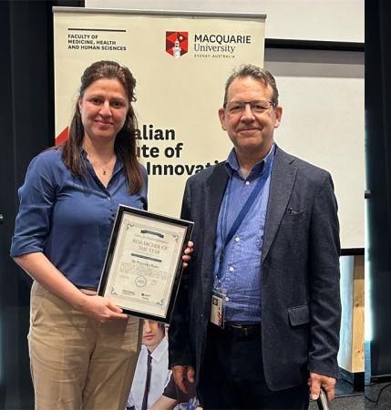
 Grace Attrill is the recipient of the Warren Meanwell Memorial Award. CD8+ T cells are immune cells which can target and destroy cancer cells, and many immune therapies – such as anti-PD-1 therapy – work by boosting these cells. Immunotherapy is not 100% effective for all melanoma patients, and researchers are currently working to figure out why. Intriguing new research has suggested that many of the CD8+ T cells in tumours do not target melanoma cells. Understanding how this occurs could be vital to improving melanoma therapies and reducing treatment side-effects. However, with current technologies it’s difficult to differentiate between the CD8+ T cells that target melanoma – the ‘melanoma-specifics’ – and those that do not – the ‘bystanders’.
Our study will combine multiple cutting-edge technologies to generate gene expression signatures that distinguish melanoma-specific and bystander CD8+ T cells in the tumours and blood of metastatic melanoma patients treated with anti-PD-1 therapy. With these signatures, we will learn more about melanoma-specific and bystander CD8+ T cell functions and their roles in immunotherapy response and resistance. It is hoped that generating these CD8+ T cell profiles for melanoma patients will lead to more personalised and effective therapies.
 My research involves investigating the utility of a scarless biopsy technique which collects skin samples using adhesive tape. Proteins are extracted from the collected skin samples to identify a protein signature which will diagnose melanoma in suspicious moles. We are recruiting patients undergoing full body photography through the Australian Centre of Excellence in Melanoma Imaging and Diagnosis (ACEMID) to provide comprehensive imaging and histopathology together with the scarless biopsy.
We aim to develop a diagnostic approach which will reduce the number of removed moles with no malignant potential, improve detection of melanoma, and provide an accessible tool for patients in rural and remote communities.
 The use of immunotherapy has revolutionised the melanoma treatment landscape in the last decade. End-stage melanoma patients now experience improved survival, but many suffer from debilitating, potentially severe grade immune-related side adverse events (irAEs) that require hospitalisation, potential treatment disruption and may be fatal. Biomarkers in the blood of patients that can predict treatment-related irAEs do not currently exist but are critical for the monitoring and prevention of these severe side effects. Based on our previous data, we believe that autoantibodies, markers of the immune system, may serve as predictive biomarkers of severe side effects.
We recently identified and will now validate a shortlist of autoantibodies that may be predictive of severe side effects and these may prove valuable for the development of a blood test that can improve the prediction and early detection of immunotherapy side effects in melanoma patients. In the future, such a blood test has the potential to aid in clinical decision-making with regard to treatment options. This will especially be important in coming years since these treatments are now considered for use in much earlier tumour stages (stage 2 onwards) and the predicted risk of irAE development will be an important consideration for the use of these treatments at early disease stages.
 Melanoma is Australia’s 3rd most common cancer. Thus, prevention and early detection is key to ensuring better health outcomes for Australians. While population-wide screening is not economically feasible for melanoma, targeting screening for high-risk individuals is a potentially viable solution. Currently, traditional risk factors (i.e. sun damage, skin colour, etc) are used to identify individuals at increased risk of melanoma. However, including genetic risk factors (called polygenic risk factors) to calculate an overall risk estimate can more accurately identify those at highest risk of melanoma. This new test is called integrated risk.
This study aims to better understand the additional value of polygenic risk information in melanoma risk assessments, beyond what traditional risk factors provide, including psychosocial (i.e. perceived personal utility, distress, anxiety, and empowerment) and behavioural outcomes (i.e. sun-safety and screening adherence). Specifically, the study aims to evaluate differences in between individuals that receive information booklets with melanoma risk-estimates based on integrated scores (polygenic risk inclusive) versus those who receive melanoma risk-estimates based on traditional risk factors at one-month follow up.
 Immunosuppressed patients are at significantly higher risk of melanoma, have worse disease-specific outcomes, and are in growing numbers in Australia. However, there is limited research investigating the biology of melanoma in this group.
The aim of my PhD project involves defining the composition of the tumour microenvironment in melanoma from immunosuppressed and immunocompetent patients using a novel tissue imaging technology called Imaging Mass Cytometry (IMC) and investigating potential disease biomarkers. IMC permits the characterisation of hundreds of cell populations in tumours, and their location and interactions with one another. Currently, there are no population-specific biomarkers predicting clinical behaviour or pathologic features of melanoma in immunosuppressed patients. Our selected cohort has long-term outcome data (including treatment responses) available, permitting the investigation of relevant clinicopathologic biomarkers.
This is one of the first studies to research the biology of melanoma in immunosuppressed patients. Hopefully, findings from this project will lead to increased understanding of melanoma in this high-risk population, and ultimately to improved melanoma outcomes.
 The Lentigo Maligna Spectrum Project aims to answer a crucial clinical question for melanoma management: how can we differentiate a melanoma in its very early stages from an invasive melanoma?
Lentigo Maligna represents the most prevalent form of melanoma in situ in Australia with an incidence rising rapidly.
There is an urgent need to improve the diagnostic accuracy of the Lentigo Maligna and its invasive variant, Lentigo Maligna Melanoma, in order to establish when it is safe to treat it with non-surgical modalities versus when surgery is mandatory, and which surgical margins are necessary.

This project has the potential to improve the survival of the thousands of Australians diagnosed with early stage melanoma each year.
Early-stage (stage 1-11) melanoma accounts for the largest proportion of new melanoma diagnoses, with a total number higher than all other stages combined. We aim to understand the mechanisms involved in melanoma recurrence after surgery by performing a comprehensive analysis of the clinical, pathological, molecular and genomic characteristics of early-stage melanoma with extreme clinical outcomes, such as very thin melanomas (<1mm) that recur rapidly after surgery.
We will harness new, cutting-edge technology such as spatial transcriptomics to create an architectural map of cells both within and surrounding a tumour.
Our project shifts the focus from treatment of advanced disease to prevention of advanced disease, upholding the age-old adage of prevention is better than cure.
 The aim of this project is to evaluate immune biomarkers in 50 patient melanoma-invaded regional lymph nodes and site-matched and other lymph nodes without metastases. While prior work has focused heavily on T lymphocytes, we will primarily aim to phenotype Natural Killer (NK) cells and understand the interactions between NKs, type 1 dendritic cells and CD8 T cells, as these cells were shown to be highly relevant to immune control of primary melanomas. Studies in patient lymph nodes have suggested positive associations between NK cell density and patient longevity, but this has been insufficiently investigated.
Metastatic melanoma is a significant clinical problem, and the success of immune-based therapies relies on a functioning anti-tumour immune response. The lymph nodes are both control centres of locoregional anti-melanoma immune responses, and the first place melanoma spreads to in most patients.
We need to understand how melanomas interact with their draining lymph nodes better, to identify immune control points that can be manipulated for therapeutic benefit.

Exhaustion of T cells is an important cause of immunotherapy failure. Our studies have demonstrated that nicotinamide can prevent and reverse the T exhaustion state in vitro. We would like to test whether this also occurs in vivo, using a melanoma model sensitive to immunotherapy with antibodies blocking immune inhibitory receptors on T cells.
We postulate that by preventing T cell exhaustion, nicotinamide can synergize with immune checkpoint blockade. This grant will be used test this hypothesis.
 The microbes in our gut (microbiome) influence immune processes throughout the body. This includes how patients respond to immunotherapies. These therapies aim to reactivate a patient’s own immune system to recognise and kill tumour cells. However, still, nearly 50% of patients with advanced melanoma die due to resistance. Furthermore, concurrent inflammatory side effects frequently cause severe morbidities, sometimes resulting in patients having to cease therapy.
My research looks at the role of the gut microbiome during immunotherapy. Specifically, how diet and intestinal microbes influence the efficacy and safety of treatment.
My research looks at the role of the gut microbiome during immunotherapy. Specifically, how diet and intestinal microbes influence the efficacy and safety of treatment. The AMRF has supported an animal study designed to complement the existing and on-going clinical investigation in metastatic melanoma patients, involving the analysis of microbiome sequencing data as well as matched immune profiles, metabolites and nutritional patterns. This work involved using a mouse model to test whether dietary changes can beneficially alter microbiomes in a short timeframe and whether this alters both treatment responsiveness and the susceptibility to inflammatory toxicities during immunotherapy. Understanding the interactions between diet, the microbiome and the immune system will inform the feasibility and design of dietary interventions in the clinic.
The details of the studies are outlined below. COVID led to extensive delays in completing these studies. The pilot study was successful and allowed us to establish a model to study subclinical toxicities. The expanded diet study is planned to be completed by the end of this week (10th June). I am now in the analysis phase and experiments are being conducted on samples that have been collected including microbiome sequencing, metabolite analysis and histology. I hope to include this work in manuscripts aiming to be submitted by the end of this year and I am now also planning follow up studies based on some of the promising results.
AMRF 2021 research grant recipient and PhD Candidate Rebecca Simpson has released her research findings – How diet patterns can be linked to improvements in immunotherapy.
Rebecca’s work was featured on Channel 9. To see the full report click here.
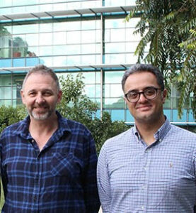 Dr Faridi has developed “HybridFinder” technology to identify “spliced peptides”, a new class targets of cancer immunotherapy. He has shown spliced peptides are abundant in melanoma, are highly immunogenic and have a high potential for clinical studies. In this project and with the help from the AMRF, Dr Faridi will work alongside Prof Anthony Purcell (Monash University) to evaluate the presence of a set of immunogenic spliced peptides in melanoma tumour biopsies.
This research will contribute towards the development of new immunotherapy strategies for melanoma treatment.
We used Peptide PCR technology and confirmed the presence of 10 spliced peptides derived from melanoma antigen in tumour biopsies from melanoma patients. In addition, validated peptides have been used in 1 patines in a clinical study in collaboration with Prof Gerry Linette (the University of Pennsylvania, The Parker Institute for Cancer Immunotherapy) and A/Prof Andreas Behren (Olivia Newton-John Cancer Research Institute) on dendritic cell-based vaccines and cancer immunotherapy (ClinicalTrials.gov Identifier: NCT03092453 “Dendritic Cell Vaccination in Patients with Advanced Melanoma”). The patient received the vaccine, and his immune system responded to our antigens. We are doing a follow-up on this patient and exploring the next patient.
Other outcomes include – I have started my independent lab at the School of Clinical Sciences at Monash University, which this grant was beneficial for this process.
Cutaneous melanoma is the most aggressive type of skin cancer that is responsible for more than 80% of skin cancer-related deaths. The incidence of cutaneous melanoma, especially thin (< 1.0 mm) melanomas, continues to increase nationally and globally. Although thick (≥ 4.0 mm) melanomas are correlated with a worse prognosis, thin melanomas account for the majority of melanoma deaths due to the high volume of the disease. Therefore, there is currently an urgent need to identify melanoma-associated prognostic biomarkers for the effective monitoring of patients that are predicted to have an increased risk of tumour progression. This will enable timely therapeutic intervention and ultimately decrease melanoma attributed morbidity and mortality.
The utilisation of autoantibodies as melanoma-associated biomarkers is a promising avenue towards personalised medicine. Autoantibodies are generated by the adaptive immune system in cancer patients towards autologous antigens and may provide biological information of the tumour. Additionally, autoantibodies have desirable biomarker properties such as persistent concentrations and long half-lives due to a limited proteolysis and clearance from the blood.
Immunotherapy, a form of treatment that aims to harness and boost a patients’ own immune system, is the current standard of care for patients with advanced metastatic melanoma. This treatment regimen is leading the way in increasing survival rates in patients whose melanoma has metastasised to other sites around their body. While these therapies have bettered outcomes for many patients and show promise in achieving long term control of their disease, there are still a large number of patients who do not respond to these treatments.
We are continually developing our understanding of the importance of tailored treatments that are as personalised as possible to achieve better long term outcomes for individual patients. We are now aware that the area of the body that melanoma spreads to, i.e. the liver, the lung, etc. can have a significant impact on the chances of a patient responding to therapy.
Specifically, we know patients whose melanoma has spread to their liver tend to respond worse than those without liver metastases.
 Uveal melanoma is the most common cancer of the eye in adults.
Approximately 8.6 per million per year are diagnosed with the disease in Australia. Although rare, about 50% will have spread of the disease from the eye to other organs and sites within the body, and once this occurs approximately 92% of people will die within two years. Unlike cutaneous (skin) melanoma, there are no effective systemic therapeutic agents for control of the disease. Currently, only complete surgical removal of metastatic tumour impacts overall survival. This, however, requires early detection of the disease via radiological scans, which can be unspecific and have limited sensitivity.
This project aims to develop a minimally invasive, robust and UM-specific blood test to supplement the current standard of care.
In a multicentre study (ECU, Aus; Erasmus MC, NL), we found that we could detect ctDNA in 63% of patients before (median lead time of 4.4 months) or at the diagnosis of metastatic disease. There was a positive and negative predictive value of 100% and 69%, respectively. Patients with detectable ctDNA had shorter survival, and higher levels were significantly associated with very poor outcomes. The levels of ctDNA detected were also significantly associated with disease burden measured using PET/CT, indicating ctDNAs’ use as a surrogate of total disease volume. Lastly, we monitored patients undergoing immunotherapy. We found that patients undergoing combination Ipilimumab/Nivolumab showed a one to two log reduction of ctDNA after treatment initiation, indicating some effect of the treatment on the tumour. In contrast, patients with single agent Pembrolizumab had no change in ctDNA levels.
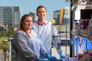 Introducing Dr Pablo Garcia Valtanen, supervisor for Ms Samantha Watson, a PhD student at University of South Australia (UniSA), who is investigating the potential for treating melanoma with a medicine that has successfully targeted oesophageal cancer cells in the laboratory. Although different, certain cancerous cells in the oesophagus and in melanoma share common traits such as the expression of disease specific molecules on their surface.
One of these molecules, is the focus of Ms Watson’s project which is trying to find new ways for treating melanoma. Her goal is to establish the potential for the use of antibodies in the clinic. This strategy has already generated positive results with oesophageal cancer models and Ms Watson now expects to replicate this success in melanoma cells.